x
-
Call Us
-
Send Email
- Dummy
Oral Cancer Exam
-
Carroll Dentistry – Procedures – Preventive Care – Oral Cancer Exam
Oral cancer is a pathologic process which begins with an asymptomatic stage during which the usual cancer signs may not be readily noticeable. This makes the oral cancer examinations performed by the dentist critically important. Oral cancers can be of varied histologic types such as teratoma, adenocarcinoma, and melanoma. The most common type of oral cancer is malignant squamous cell carcinoma. This oral cancer type usually originates in lip and mouth tissues.
There are many different places in the oral cavity and maxillofacial region in which oral cancers commonly occur, including:
- Lips
- Mouth
- Tongue
- Salivary Glands
- Oropharyngeal Region (throat)
- Gums
- Face
Reasons for oral cancer examinations
It is important to note that around 75% of oral cancers are linked with smoking, tobacco use, and excessive alcohol consumption. Dr. Carroll can provide literature and education on oral cancer prevention, making healthy lifestyle changes, and smoking cessation.
When oral cancer is diagnosed in its earliest stages, treatment is generally very effective. Any noticeable abnormalities in the tongue, gums, mouth or surrounding area should be evaluated by a health professional as quickly as possible. During the oral cancer exam, Dr. Carroll and your dental hygienist will carefully examine the maxillofacial and oral regions for signs of pathologic changes.
The following signs will be investigated during a routine oral cancer exam:
- Red patches and sores – Red patches on the floor of the mouth, the front and sides of the tongue, white or pink patches which fail to heal and slow healing sores that bleed easily can be indicative of pathologic (cancerous) changes.
- Leukoplakia – This is a hardened white or gray, slightly raised lesion that can appear anywhere inside the mouth. Leukoplakia can be cancerous, or may become cancerous if treatment is not sought.
- Lumps – Soreness, lumps or the general thickening of tissue anywhere in the throat or mouth can signal pathological problems.
Oral cancer exams, diagnosis and treatment
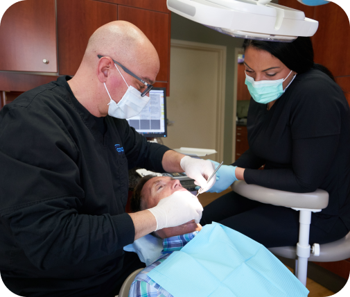
The oral cancer examination is a completely painless process. During the visual part of the examination, Dr. Carroll will look for abnormalities and feel the face, glands, and neck for unusual bumps. Lasers that can highlight pathologic changes are also a wonderful tool for oral cancer screening. The laser can “look” below the surface for abnormal signs and lesions which would be invisible to the naked eye.
If abnormalities, lesions, leukoplakia or lumps are apparent, the dentist will implement a diagnostic impression and treatment plan. In the event that the initial treatment plan is ineffective, a biopsy of the area will be performed. The biopsy includes a clinical evaluation which will identify the precise stage and grade of the oral lesion.
Oral cancer is deemed to be present when the basement membrane of the epithelium has been broken. Malignant types of cancer can readily spread to other places in the oral and maxillofacial regions, posing additional secondary threats. Treatment methods vary according to the precise diagnosis, but may include excision, radiation therapy and chemotherapy.
During bi-annual check-ups, Dr. Carroll and your hygienist will thoroughly look for changes and any lesions in the mouth, but a dedicated comprehensive oral cancer screening should be performed at least once each year.
If you have any questions or concerns about oral cancer, please ask Dr. Carroll or your dental hygienist.
Videos
Play Video
Play Video
Play Video
Play Video